
Medical review status: This article provides evidence-informed general health information. It has not been medically reviewed by a named healthcare professional.
Medical disclaimer: A lump cannot be reliably diagnosed from its firmness, location, or appearance alone. This article is educational and is not a substitute for an examination by a qualified healthcare professional.
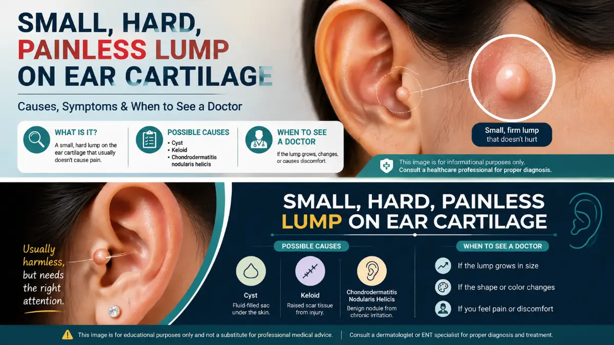
A small hard painless lump on ear cartilage is often caused by a benign skin, scar, or cartilage-related condition, but there is no single diagnosis that fits every hard ear bump.
Possibilities include a normal cartilage prominence, an epidermoid cyst, scar tissue after a piercing, an auricular pseudocyst, a gouty tophus, or another skin lesion. Chondrodermatitis nodularis helicis (CNH) is also well known for causing a small firm nodule on the helix or antihelix, although CNH is typically painful or tender, so a completely painless bump is less characteristic.
A new growth that keeps enlarging, becomes crusted or ulcerated, bleeds, changes appearance, or fails to heal deserves medical assessment because skin cancers can also develop on the sun-exposed ear.
Key Takeaways
- A painless hard bump is a description, not a diagnosis.
- Previous ear piercing or injury makes a raised scar or keloid more plausible.
- A smooth lump underneath the skin may represent an epidermoid cyst.
- A painless swelling within the upper ear cartilage can sometimes be an auricular pseudocyst.
- CNH usually causes tenderness or pain, making it less typical when the lump is completely painless.
- People with longstanding gout can develop hard, sometimes painless urate deposits called tophi on the ears.
- Persistent growth, crusting, bleeding, ulceration, or enlargement should not be ignored.
- Do not cut, puncture, squeeze, or repeatedly pick at an unexplained hard ear lump.
What Does a Small Hard Painless Lump on Ear Cartilage Usually Mean?
The cartilage-containing portion of the visible ear is called the auricle or pinna. It includes the helix, antihelix, concha, tragus, and other folds. The earlobe is different because it does not contain cartilage.
This distinction matters. A bump actually sitting on the upper or middle ear cartilage has a somewhat different group of possible causes than a lump inside the soft earlobe or behind the ear.
The following clues are useful, but none can confirm a diagnosis by themselves.
Also Read: Knee Cartilage: Damage, Symptoms, Treatment, Exercises & Recovery
| Feature | Conditions it may suggest |
| Hard and unchanged for years | Normal cartilage prominence or longstanding benign lesion |
| Round lump beneath skin | Epidermoid cyst |
| Appeared after piercing or injury | Keloid or other scar tissue |
| Smooth painless swelling in upper pinna | Auricular pseudocyst |
| Firm helix/antihelix nodule with tenderness or crust | CNH |
| Hard white/yellowish nodules with history of gout | Gouty tophi |
| Growing, crusting, ulcerating or bleeding lesion | Requires assessment for skin cancer or another skin disorder |
1. Normal Ear Cartilage or an Anatomical Variant
Not every hard prominence is an abnormal growth.
The outer ear naturally contains several firm cartilage ridges. Normal ear anatomy also varies substantially between people and even between a person’s left and right ears.
One recognized normal variant is Darwin’s tubercle, a small prominence along the helix. It has no known harmful function or disease association.
A normal anatomical prominence becomes more likely when the area:
- has apparently been present for many years,
- feels identical to the surrounding cartilage,
- has normal skin covering it,
- does not grow or change,
- has no crusting, bleeding, discharge, redness, or tenderness.
A genuinely new lump should not automatically be assumed to be normal anatomy.
2. Epidermoid Cyst
An epidermoid cyst is a benign cyst containing keratin beneath the skin. These cysts often appear as round nodules and may have a tiny central opening or punctum.
They can remain painless when they are not inflamed. They often feel firm but tend to be more mobile than the cartilage underneath them. Inflamed cysts can become red, swollen, tender, and more noticeable. An epidermoid cyst develops in the skin rather than being a true growth of the cartilage. Avoid squeezing one. Trauma can trigger inflammation, rupture, infection, or scarring.
3. Keloid or Raised Scar After an Ear Piercing
If the bump formed at or near an old piercing, scar tissue becomes an important possibility.
A keloid is an overgrowth of scar tissue that extends beyond the boundaries of the original injury. Ear piercings are a recognized trigger. Keloids may be firm or rubbery and can grow gradually over months. Some itch or hurt, while others cause few symptoms.
A piercing-related lump should therefore be evaluated in the context of:
- exactly where the piercing entered the ear,
- when the lump first appeared,
- whether it continues to enlarge,
- whether similar scars have occurred previously.
Dermatologists may use treatments such as corticosteroid injections, pressure therapy, silicone therapy, cryotherapy, or combined surgical approaches depending on the individual keloid.
4. Auricular Pseudocyst
An auricular pseudocyst is an uncommon benign swelling caused by a fluid-containing space within the ear cartilage.
It typically develops in the upper part of the pinna and is usually painless and non-inflammatory. This can fit the “painless ear cartilage lump” description well, but pseudocysts generally have a swelling or slightly fluctuant quality rather than feeling like a tiny piece of solid bone.
A clinician may distinguish a pseudocyst from a cyst, hematoma, cartilage abnormality, or other lesion during examination. It should not be punctured at home because recurrence, infection, and cartilage damage are possible concerns.
5. Chondrodermatitis Nodularis Helicis
Chondrodermatitis nodularis helicis, usually shortened to CNH, is a benign inflammatory condition affecting the skin and cartilage of the helix or antihelix. It characteristically produces a small, firm nodule, often with a central crust. Repeated pressure from sleeping on the affected ear is considered an important contributing factor. Earphones, hearing aids, cold exposure, trauma, and sun exposure may also contribute.
However, there is one particularly important distinction for this search:
CNH is characteristically painful or tender.
Therefore, if you have a completely small hard painless lump on ear cartilage, CNH is possible but is not the most textbook presentation.
Persistent CNH-like lesions sometimes require biopsy because CNH can resemble squamous cell carcinoma and other lesions.
6. Gouty Tophus
People with longstanding gout may develop tophi, which are collections of monosodium urate crystals. Tophi can occur under the skin of the ears and may feel hard. Importantly, they are often painless unless inflamed.
This possibility is more relevant when a person already has:
- diagnosed gout,
- previous attacks of sudden painful joint inflammation,
- similar firm nodules around the fingers, toes, elbows, or Achilles tendon,
- multiple white, cream-colored, or firm nodules.
An isolated ear bump does not by itself establish a diagnosis of gout.
7. Could a Painless Ear Lump Be Skin Cancer?
Most small ear lumps are not automatically cancerous. Still, cancer cannot be ruled out simply because a lesion is painless. The ears receive substantial ultraviolet exposure. Both basal cell carcinoma and squamous cell carcinoma commonly develop on sun-exposed areas of the head and neck, including the ears. Squamous cell carcinoma can sometimes resemble a firm, crusty or enlarging growth.
Have a clinician examine a lesion if you notice:
- progressive enlargement,
- repeated crusting,
- a sore that does not heal,
- spontaneous bleeding,
- ulceration,
- significant color or surface changes,
- irregular borders,
- persistent scaling,
- destruction or distortion of the surrounding ear.
Appearance alone is not enough to distinguish benign conditions such as CNH from some skin cancers. A dermatologist may recommend a biopsy when the diagnosis is uncertain.
Read More: Human Torso Anatomy: A Complete Guide to the Body’s Core Structure
Location Provides Useful Clues
Before searching for a diagnosis, determine exactly where the lump is located.
Outer rim of the ear – helix
Possible considerations include:
- normal cartilage prominence,
- CNH,
- skin cyst,
- gouty tophus,
- sun-related skin lesions.
Inner cartilage ridge – antihelix
CNH and auricular pseudocysts can occur in this region, although their typical texture and symptoms differ.
At an old cartilage piercing
Keloid or other scar tissue becomes more relevant.
Soft earlobe
The earlobe does not contain cartilage. A lump there therefore falls outside the exact meaning of a lump “on ear cartilage.” Cysts and piercing-related scars are common considerations.
Behind the ear
A bump behind the ear is also a different problem. The possible causes can include skin cysts, lymph nodes, normal underlying bone, infection, and other conditions.
A Simple Self-Check Before Your Appointment
Do not try to diagnose the bump yourself, but these observations can help a clinician.
Ask:
- When did I first notice it?
- Has its size changed?
- Is it actually on the ear cartilage, under the skin, in the earlobe, or behind the ear?
- Does it move separately from the cartilage?
- Is the surface smooth, rough, scaly, crusted, or ulcerated?
- Has it ever bled or drained?
- Was this area previously pierced or injured?
- Do headphones, helmets, hearing aids, or sleeping positions repeatedly press on it?
- Do I have gout or similar lumps elsewhere?
- Has the surrounding skin become red, warm, swollen, or painful?
Taking a clear photograph now can also help you objectively determine whether it changes over time.
When Should You See a Doctor?
Arrange an examination if the lump is new and persistent, unexplained, growing, or changing.
Evaluation is particularly important when there is:
- bleeding,
- crusting or ulceration,
- rapid enlargement,
- a non-healing sore,
- significant color change,
- distortion of the ear,
- recurring discharge,
- a history of substantial sun exposure combined with a changing lesion.
Seek prompt medical care for signs of infection
A cartilage piercing or ear lesion accompanied by increasing redness, warmth, swelling, pus, worsening pain, fever, chills, or feeling generally unwell requires faster assessment. The NHS advises seeking medical care for suspected infected piercings because complications can become serious.
How Doctors Diagnose an Ear Cartilage Lump
Most evaluations begin with history and physical examination.
A clinician may assess:
- exact anatomical location,
- size and firmness,
- mobility,
- surface texture,
- pigmentation,
- surrounding inflammation,
- piercing or trauma history,
- sun exposure,
- whether the lesion is growing.
Dermatologists can use dermoscopy to examine surface structures more closely. If the lesion could represent CNH, skin cancer, or another uncertain skin growth, a biopsy may be needed. DermNet notes that inflammatory and neoplastic lesions of the ear can look similar and that careful examination and sometimes biopsy are necessary for accurate diagnosis.
Imaging is generally not required for every tiny superficial skin bump but may be considered when a clinician suspects a deeper cartilage or surrounding structural problem.
What Treatment Might Be Needed?
Treatment depends entirely on what the lump actually is.
Read More: Emergency Dentist: What to Do When You Need Immediate Dental Care
| Cause | Typical management |
| Normal cartilage variant | Usually none |
| Uninflamed epidermoid cyst | Observation or medical removal if needed |
| Keloid | Dermatologic scar treatment, often combination therapy |
| Auricular pseudocyst | ENT/dermatology treatment such as drainage plus compression or another procedure |
| CNH | Pressure reduction and clinician-directed medical or procedural treatment |
| Gouty tophus | Treatment of underlying gout |
| Suspicious skin lesion | Dermatology evaluation and biopsy/removal when indicated |
| Infection | Appropriate medical treatment based on severity and cause |
What Not to Do
Until you know what the lump is:
- do not pierce it with a needle,
- do not cut it,
- do not repeatedly squeeze it,
- do not scrape off recurring crusts,
- do not apply strong acids or wart-removal products without a diagnosis,
- do not assume “painless” automatically means harmless.
Manipulation can create inflammation, infection, bleeding, or scarring and can make the original lesion more difficult to evaluate.
Which Doctor Should You See?
A primary-care clinician can perform the initial examination and refer when appropriate. A dermatologist is particularly appropriate for a bump involving the skin surface, pigmentation, crusting, bleeding, a suspected cyst or keloid, or concern about skin cancer. An ENT specialist (otolaryngologist) may be especially useful when the swelling appears to arise within the cartilage itself, affects ear structure, involves the ear canal, or may represent a pseudocyst or another auricular disorder.
Frequently Asked Questions
Is a small hard painless lump on ear cartilage usually serious?
Not necessarily. Benign causes include normal cartilage anatomy, cysts, scar tissue, pseudocysts, and other non-cancerous lesions. However, persistence, growth, bleeding, ulceration, or surface changes warrant medical evaluation.
Is CNH always painful?
Pain and tenderness are classic features of chondrodermatitis nodularis helicis. A completely painless lesion is therefore less typical for CNH, although symptoms alone cannot exclude a diagnosis.
Can an ear piercing leave a hard painless bump?
Yes. Piercing injuries can trigger keloids or other raised scars. Ear piercing is a recognized cause of keloid formation.
Can a cyst feel hard on the ear?
Yes. Epidermoid cysts may feel firm, especially when small. They are usually located beneath the skin rather than being a true cartilage growth.
Should I pop a lump on my ear cartilage?
No. An unexplained cartilage or skin lump should not be punctured or squeezed. Doing so can cause inflammation, infection, bleeding, scarring, or cartilage injury.
Could gout cause a painless lump on the ear?
Yes. Gouty tophi can occur beneath the skin of the ears and are normally painless unless they become inflamed. This is usually considered in the context of gout or other tophi.
How long should I wait before having an ear lump checked?
There is no universal safe waiting period for every lesion. A new lump that persists, enlarges, changes, repeatedly crusts or bleeds, or has no clear benign explanation should be examined rather than monitored indefinitely.
Can you tell what an ear lump is from a photograph?
Sometimes photographs provide useful clues, but many ear lesions overlap in appearance. A physical examination-and occasionally dermoscopy or biopsy-is necessary when the diagnosis remains uncertain.